* Figures based on tendon pathology examinations undertaken at Barossa Equine. Please enquire for further data.
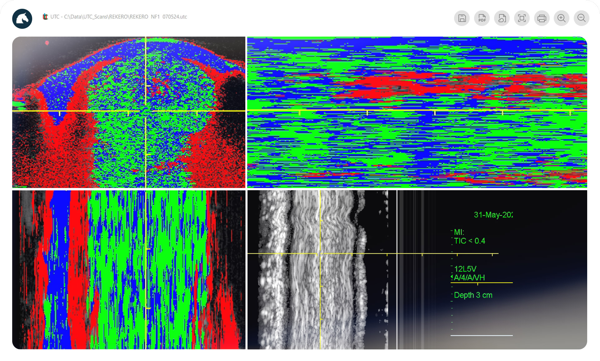
Ultrasound Tissue Characterisation (UTC) is an advanced imaging technology that provides unique insights into tendon and ligament health. Unlike traditional ultrasound, which offers only a structural view, UTC uses specialised algorithms to analyse tissue composition at a microscopic level. It scans the tendon in high-resolution slices, capturing the alignment, density, and integrity of individual fibres.
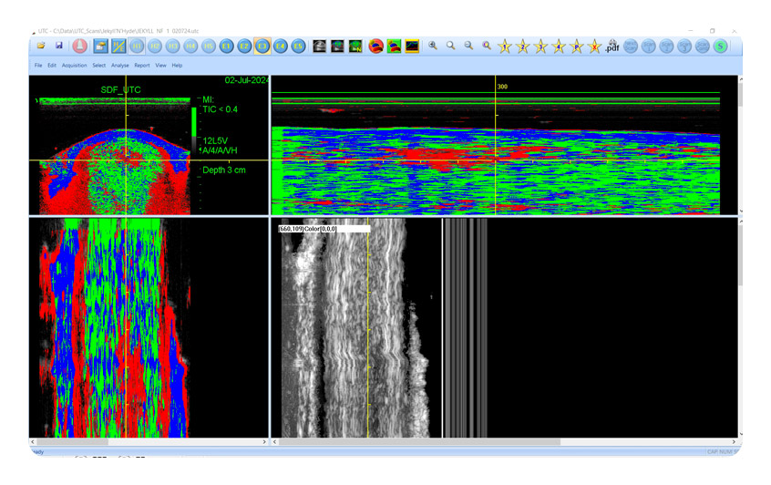
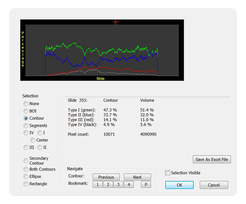
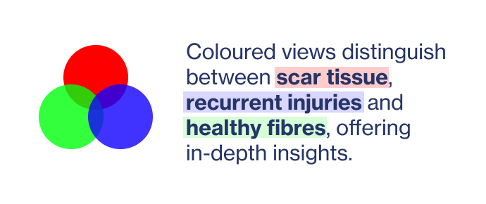
This process creates a colour-coded, three-dimensional image that distinguishes healthy fibres from those that are swollen, disorganised, or damaged. These images allow veterinarians to monitor the healing process in real-time and adjust treatment as tissue changes.
By showing what’s happening within the tendon, UTC makes it possible to create tailored rehabilitation plans that adapt as the tendon heals, helping to prevent re-injury and ensuring more effective recovery. Widely trusted in elite sports, UTC technology brings the same high standard of care to equine rehabilitation, supporting long-term health and performance.
The table below indicates the differences between traditional ultrasound and UTC.
Uses high-frequency sound waves to create images of soft tissue.
Image Quality
Produces two-dimensional, grayscale images that show the structure of tendons. It can detect tendon tears, inflammation, and changes in tendon shape.
Common Use
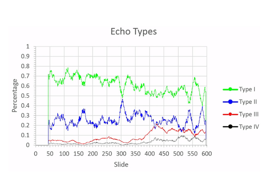
UTC is particularly useful for tracking the quality of tendon tissue during rehabilitation, helping to determine if tissue is remodeling appropriately.
Limitations / Advantages
Limited by image resolution; it cannot differentiate subtle changes in tissue quality, making it harder to evaluate tissue remodeling during rehabilitation.
The table below indicates the differences between traditional ultrasound and UTC.
Uses a more sophisticated algorithm to analyse sound waves as they pass through tissues, offering a quantitative assessment.
Produces a high-resolution, color-coded image showing four distinct tissue types based on their structural integrity:
intact, fibrillar, swollen, or damaged.
Produces two-dimensional, grayscale images that show the structure of tendons. It can detect tendon tears, inflammation, and changes in tendon shape.
Common Use
UTC is particularly useful for tracking the quality of tendon tissue during rehabilitation, helping to determine if tissue is remodeling appropriately.
Limitations / Advantages
With traditional/grey scale ultrasound, rehabilitation programs are developed on time from injury and ultrasound assessment of the tendons fibre alignment in a two dimensional plane. While this is satisfactory to a degree, its weakness is the variable rate of change through the healing stages. It may miss the crucial, but small window of optimum time for developing fibre alignment. UTC technology gives more qualifying and quantifying information on tendon structure throughout the healing stages.
Traditional ultrasound gives us partial information. Colourised zones of UTC supply information about whether the tendon is intact, fibrillar, swollen or damaged.
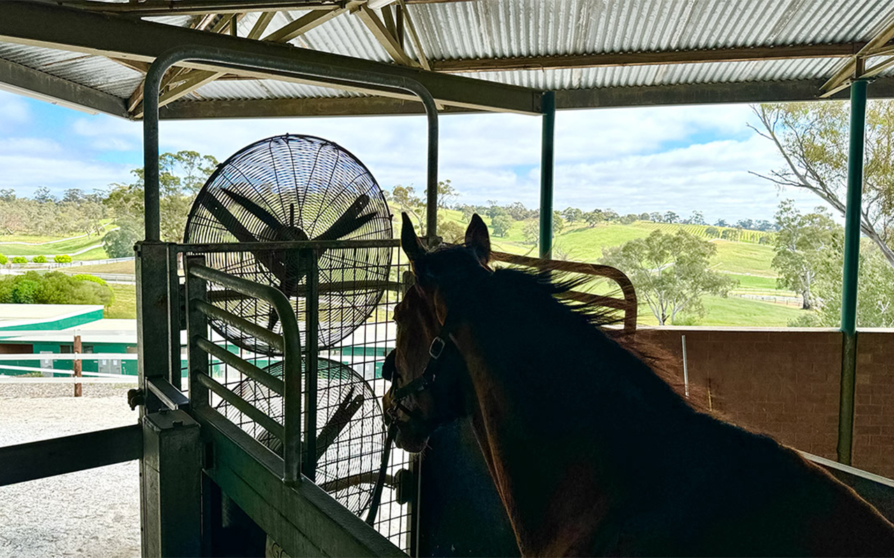
UTC technology first proved its value in pro-sport, used in professional European Football Leagues and in the AFL to monitor and protect athletes’ tendon health. Developed to support human performance, UTC’s success in high-stake environments led to its adoption in equine care.
This technology is now available in the equine industry, using UTC to provide the same level of precision and insight trusted by top athletes.